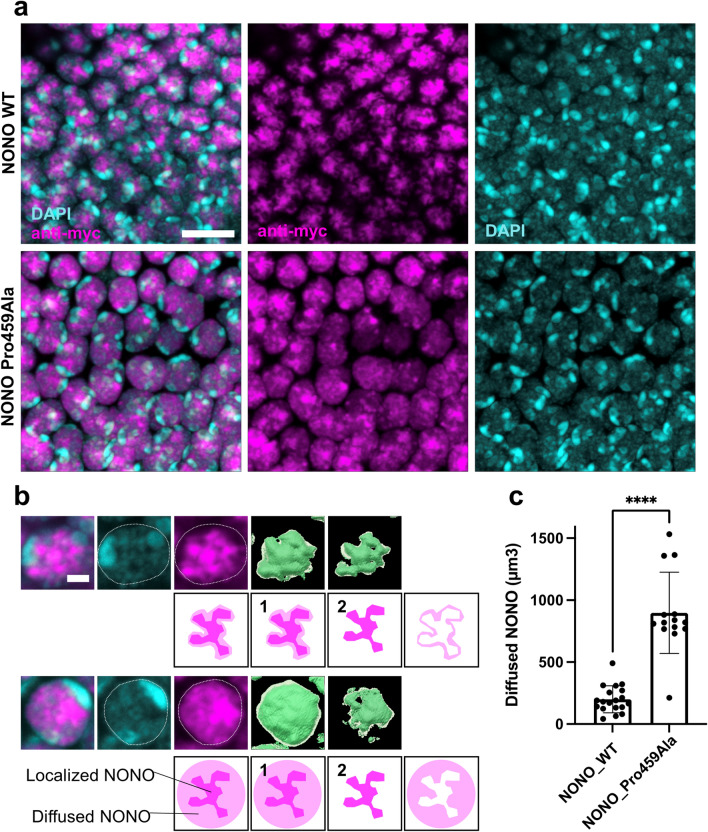
Figure 3.
Localization pattern of NONO variant in Drosophila neuron. (a) Cell bodies of Kenyon cells in Drosophila brain. Wild-type NONO and Pro459Ala NONO are expressed. The localization pattern of Myc-NONO (magenta) was stained with anti-Myc antibody using immunohistochemistry. The nuclei were visualized using DAPI (cyan). Scale bar = 5 µm. (b) Comparative localization patterns of NONO variants in a Kenyon cell. Myc-NONO is shown by anti-myc antibody and the nucleus is visualized using DAPI. NONO is strongly localized in the weak region of DAPI. The Imaris image analysis software extracted (1) Myc signals, including the strong and weak signals, and (2) only strong Myc signals. They were then converted into numerical form as volumes (µm3). Diffuse signals in the nucleus were then calculated by subtracting (2) from (1). Scale bar = 1 µm. (c) Quantification results of the diffuse signals in the nucleus (µm3) in Myc-NONO WT (n = 19) and Myc-NONO Pro459Ala (n = 14). Data represent the mean ± SD. Statistical comparisons were conducted using a nonparametric Mann–Whitney test. ***P < 0.0001.

