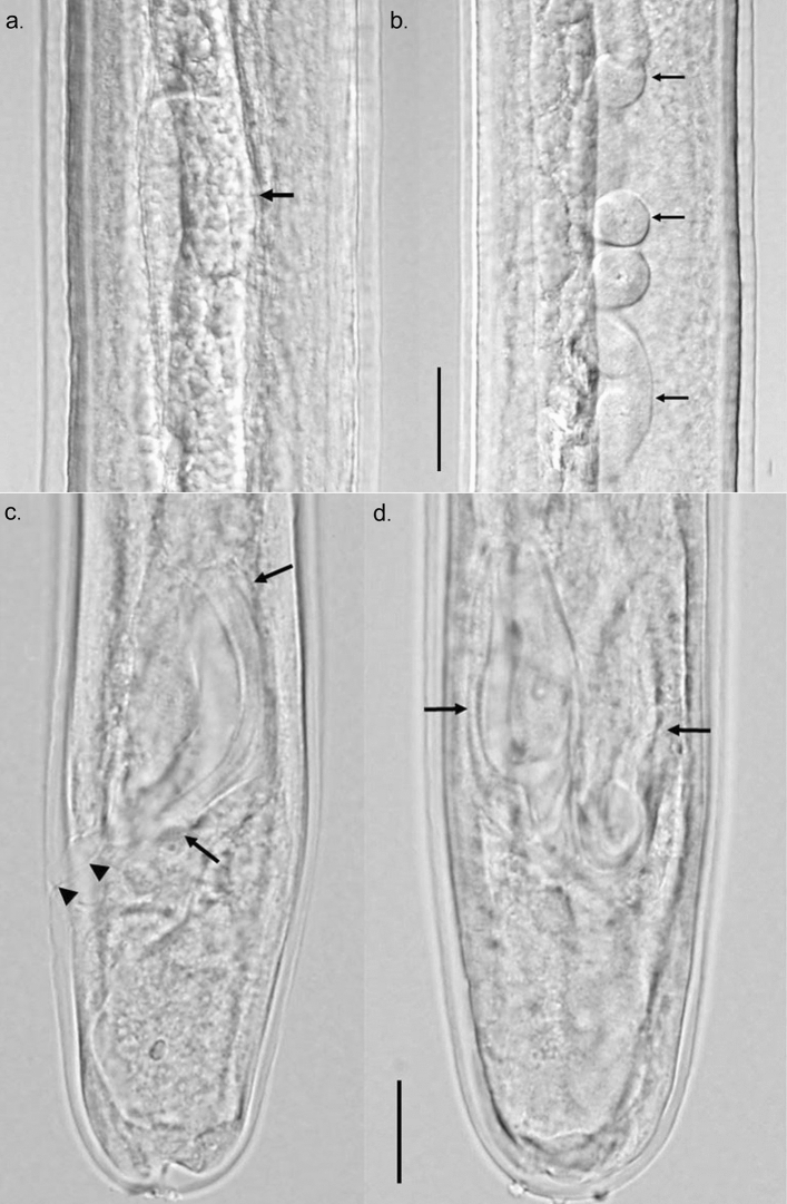
Figure 3.
Differential Interference Contrast microscopy image of male larva recovered from NSG mice at 15 weeks post infection. Composite image of testis and reproductive tube. (a) C-shaped testis (arrow) at about midbody. (b) Highly coiled male reproductive tube (arrows) in posterior third of body. Scale bar 20 µm. Composite image of posterior end of male larva showing developing spicules. (c) Tail and one spicule (arrows) in lateral view showing size, shape and cuticularization. Cloaca and anal opening (arrow heads) also visible. (d) Dorso-ventral view showing both developing spicules (arrows). Scale bar 40 µm.

