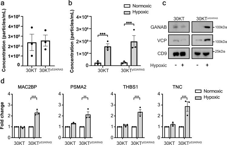
Fig. 3. The hypoxic sEV signature is derived from lung cells that are malignantly transformed.
a Nanoparticle analysis of sEVs isolated from normal or transformed HBECs shows no difference in sEV secretion. b Hypoxia significantly increases sEV secretion in HBEC lines independent of the presence of oncogenic manipulations. c Western blot of of sEVs from normal lung or malignant HBECs demonstrates that hypoxia elevates the abundance of GANAB and VCP only in malignant HBECs sEVs. d ELISA of MAC2BP, PSMA2, THBS1 and TNC in sEVs derived from hypoxic or normoxic conditions of normal or transformed lung epithelial cells indicates that only transformed lung cells increase the abundance of all four proteins in response to hypoxia (±SEM; n = 3 independent replicates). **p < 0.01, ***p < 0.001.

