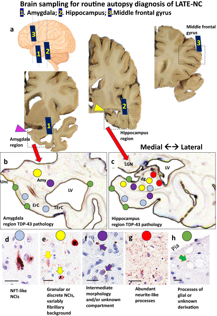
Fig. 1.
Anatomical regions of interest for tissue sampling and typical findings in routine autopsy diagnosis of LATE-NC. At autopsy, tissue portions for sampling include amygdala, mid-level hippocampus, and middle frontal gyrus. The levels of sections are shown in the cartoon form (upper left) with gross photographs of hemi-brains cut in the coronal plane (Panel a). Note that the amygdala is preferably sampled for TDP-43 immunohistochemical staining at the level of the uncus (pink arrowhead, Sect. 1), the hippocampus at the level of the lateral geniculate nucleus (yellow arrowhead, Sect. 2), and the middle frontal gyrus (Sect. 3) is sampled rather than other portions of frontal cortex. Panels b and c are representations of the amygdala region and hippocampal region, showing both the local anatomy and a cursory depiction of the subtypes of TDP-43 pathology that are generally found in those regions with corresponding colored circles in panels d-h. Panel d shows a neuronal TDP-43 + inclusion reminiscent of a neurofibrillary tangle. Panel e depicts a different TDP-43 pathologic appearance with a granular NCI (arrow with red outline), neuronal intranuclear inclusion (arrow with blue outline), and TDP-43 + fibrillary material in the background. This pattern is reminiscent of FTLD-TDP type A. TDP-43 pathology can also be present around vascular components such as capillaries (termed Lin Bodies after Ref. [87]) or in unknown histologic compartments as shown in Panel f. In some regions, the predominant TDP-43 + pathology is fine non-tapering neurite-like processes (Panel g). A different type of TDP-43-immunoreactive cell processes can be seen in the sub-pial region, often near corpora amylacea (arrow in Panel h). Scale bars = 50 microns (d); 30 microns (e); 30 microns (f); 100 microns (g); and, 30 microns (h). Abbreviations: Amyg: amygdala proper; dg: dentate granule layer of hippocampus; ErC: entorhinal cortex; LGN: lateral geniculate nucleus; LV: lateral ventricle; NCI: TDP-43 immunoreactive neuronal cytoplasmic inclusions; NFT: neurofibrillary tangles; TErC: transentorhinal cortex; Unc: uncus

