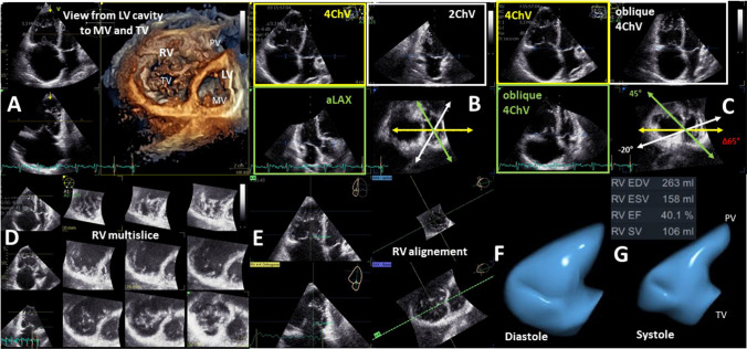
Fig. 13.
Illustration about the necessity for 3D-RV assessment -pathological conditions. A presents the enface view from the LV cavity to the MV and TV. In B the apical 3D views (4ChV, 2ChV—apical 4- and 2-chamber view; aLAX—apical long axis view) within the 3D data set are centered to the LV illustrating the standardized 4ChV by representative angular distance of 60° between aLAX, 2ChV, and 4ChV. In C oblique 4ChV are shown to illustrate that during pathological RV conditions the deviation of non-standardized 4ChVs for 2D measurements is within a Δ of 65°. In D a multisclice presentation of the 3D data set illustrate the complete acquisition of the RV volume. In E the RV alignment for RV volume determination is shown. In F the diastolic RV volume, in G the systolic RV volume is presented by a 3D volume calculation

