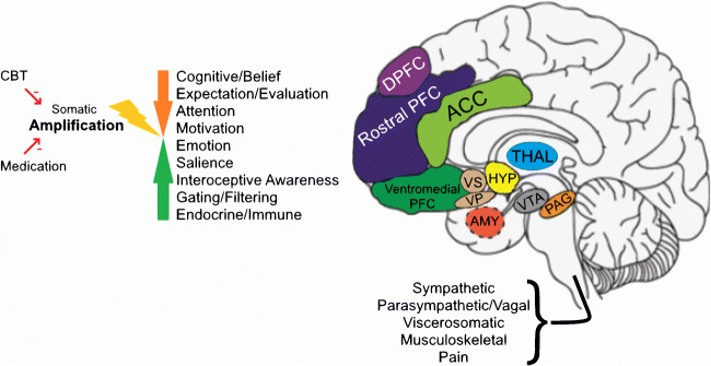
Fig. 1.
Neural circuitry of symptom perception. The figure illustrates key levels of nervous system processing of viscerosomatic and musculoskeletal experience and function. The internal organs, skin, and musculoskeletal system are innervated by a distributed neural network, with nodes and afferent and efferent connections, as well as autonomic (sympathetic and parasympathetic) modulation. The processes mediated include sensation, pain, nausea, blood flow, and movement, as well as contraction/expansion/motility-related functions. Sensory-motor signals from these viscerosomatic pathways are evaluated and modulated at more automatic, primitive, reflexic levels in the brainstem and subcortical structures, and at higher levels in specialized and associative areas of the neocortex. Evaluative and behavioral functions include affective/emotional processing and cognitive processing of bodily stimuli. The hypothalamic-pituitary axis mediates evolutionarily conserved responses, both appetitive and aversive. The insula is involved in mediating bodily awareness, the amygdala and related limbic structures are involved in triggering fight-or-flight responses, the nucleus accumbens/ventral striatum is involved in motivated behavior, the orbito-medial prefrontal cortex is involved in the integration of viscerosomatic information with socio-emotional context for decision-making and the orchestration of behavior, guided by higher order prefrontal cortex, and the anterior cingulate cortex is involved in the allocation of attentional resources and the detection and resolution of conflicting inputs and outputs. Beliefs and expectations about symptoms and illness occur at the level of multi-modal neocortex. Somatic amplification can occur through top-down and/or bottom-up amplification. Psychotherapy, pharmacotherapy, and brain stimulation target specific nodes and functions for somatic symptom and anxiety reduction. DLPFC, dorsolateral prefrontal cortex; rostral PFC, rostral prefrontal cortex; ACC, anterior cingulate cortex; ventromedial PFC, ventromedial prefrontal cortex, including orbitofrontal cortex; VS, ventral striatum; VP, ventral pallidum; HYP, hypothalamus, pituitary not shown; THAL, thalamus; AMY, amygdala; VTA, ventral tegmental area; PAG, periaqueductal gray; Insula, hippocampus, not shown.

