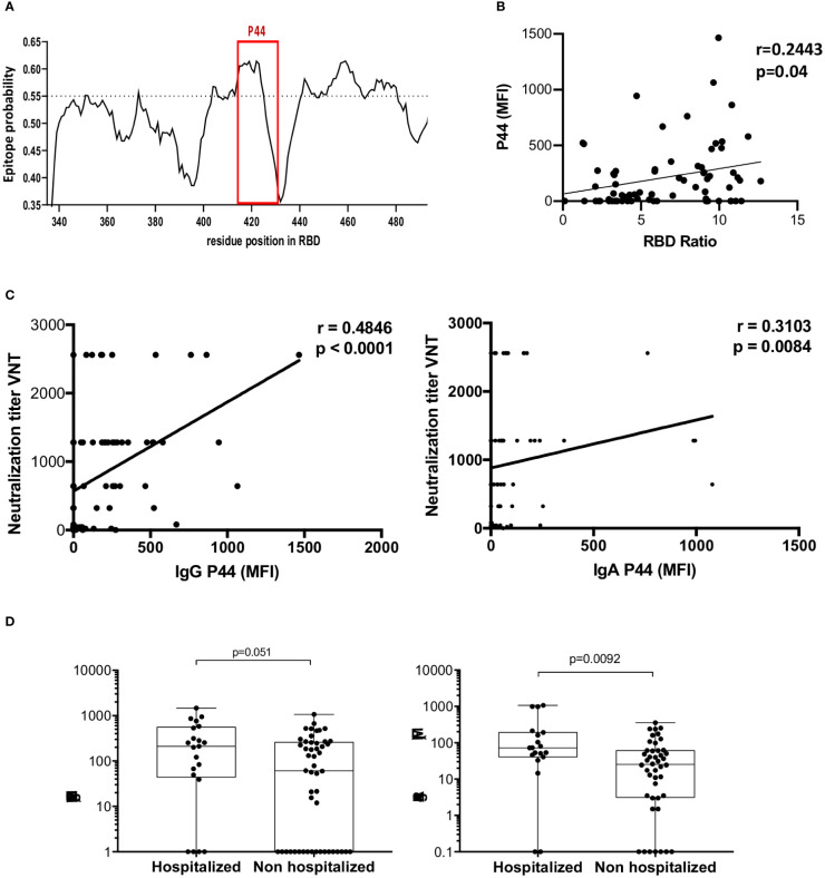
Figure 4.
Profile of P44 recognition. (A) In silico epitope prediction using BepiPred. Highlighted is the region corresponding to P44, showing a high epitope predictive value. (B) Positive correlation of P44 MFI and RBD IgG recognition in ELISA. (C) Positive correlation of IgG and IgA MFI specific for P44 and virus neutralization titers (Spearman correlation for IgG r= 0.4846, p<0.0001, 95% confidence interval 0.2769 - 0.6491, and for IgA r=0.3103, p=0.0084, 95% confidence interval 0.07602 - 0.5121). (D) Mean fluorescence intensity (MFI) of P44 antibody recognition of hospitalized compared to non-hospitalized individuals, showing higher IgA reactivity in hospitalized individuals (p=0.0092, Mann Whitney).

