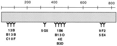
FIG. 4.
Schematic representation of the regions in VacA from H. pylori strain 60190 to which the monoclonal antibodies bind, based upon data shown in Tables 2 and 4 as well as reactivity of antibodies with mutant VacA proteins containing alanine substitution mutations (see text). The numbers represent amino acids in the mature VacA toxin from H. pylori strain 60190.

