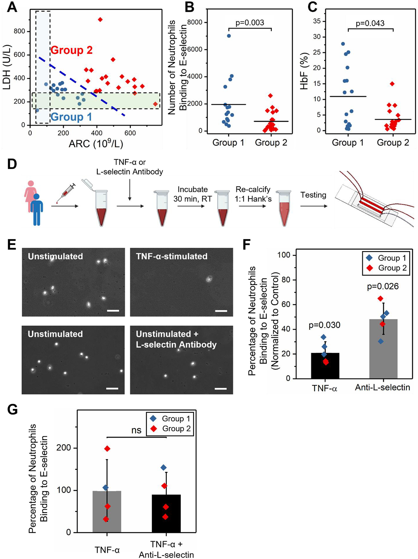
Figure 4. Neutrophil binding to E-selectin inversely correlates with vascular hemolysis in SCD and is mediated by surface L-selectin.

(A) Subjects were stratified into 2 sub-groups based on their serum LDH levels and absolute reticulocyte counts (ARCs) using the K-means clustering method. HbSS Group 2 (N = 19) samples had significantly higher serum LDH levels and ARCs but lower HbF% compared to HbSS Group 1 (N = 16). The shaded regions indicate reference ranges (normal) for LDH and ARC levels, respectively. (B) HbSS Group 2 subjects had significantly lower neutrophil binding to E-selectin compared to HbSS Group 1 subjects (P = 0.003, Mann-Whitney). (C) HbSS Group 2 subjects had significantly lower HbF% compared to HbSS Group 1 subjects (P = 0.043, Mann-Whitney). (D) Schematic representation of experimental conditions. Whole blood samples drawn from subjects with HbSS were incubated with 25 ng/mL TNF-α or 10 μg/mL anti-L-selectin antibody at room temperature (RT) for 30 min. Thereafter, blood samples were re-calcified and perfused through E-selectin microchannels. (E) Representative microscopic images of neutrophils adhered to E-selectin under the various conditions listed. Scale: 50 μm. (F) Neutrophil activation with TNF-α significantly reduced neutrophil binding to E-selectin (N = 5, P = 0.030, paired t-test). Similarly, L-selectin blocking antibody significantly reduced neutrophil binding to E-selectin (N = 5, P = 0.026, paired t-test). (G) Anti-L-selectin treatment did not reduce neutrophil binding of TNF-α stimulated neutrophils (N = 4, ns: P > 0.05).
