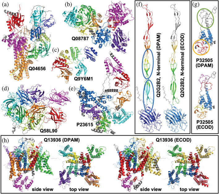
FIGURE 5.

Examples of parsed domains in AF models by DPAM. Different domains in a protein are colored from blue (or purple, N‐terminal) through green, yellow, to red (or magenta, C‐terminal). Non‐domain regions are colored in gray. (a–e) cases where DPAM domain definitions agree with ECOD definitions. (f) a case where DPAM domain definitions are not all accurate, and domains that are incorrectly split are in blue circles. (g) a case where DPAM missed some poorly modeled zinc fingers (in a green circle) and combined multiple consecutive zinc fingers (in orange circles). (H) a case where the boundaries of DPAM domains differ from ECOD domain boundaries, but the DPAM domains appear more meaningful
