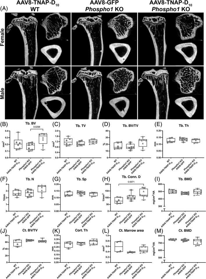
Fig. 6.

Micro‐CT analysis of tibias from Phospho1 knockout (KO) mice and wild‐type (WT) littermates. (A) 2D micro‐CT images of tibias from females and males treated with AAV8‐TNAP‐D10 or AAV8‐GFP 90 days after injection. (B–I) Quantification of trabecular bone parameters showing increased bone volume and trabecular bone connectivity in AAV8‐TNAP‐D10‐treated Phospho1 KO compared with WT and untreated Phospho1 KO mice. (J–M) quantification of cortical bone parameters showed no statistically significant difference among the groups. Statistical analysis was performed by one‐way ANOVA followed by Tukey's multiple comparison test. Differences were statistically significant at *p < 0.05 (p = 0.0298) and **p < 0.01 (p = 0.0071).
