Fig. 3.
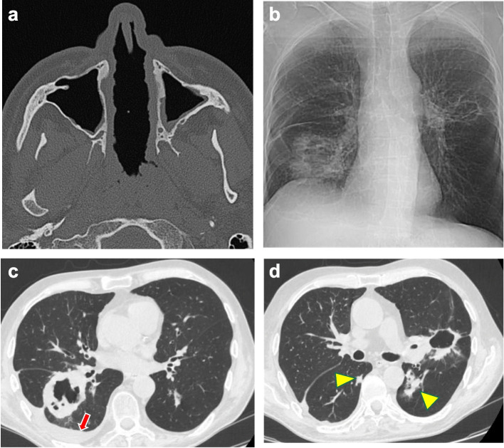
a) Necrotizing perforation of the nasal septum (patient 16 of Table 1). Axial CT demonstrates the nearly complete absence of the nasal septum. The mucosa appears thickened and nodular. b) Chest X-ray (patient 11 of Table 1), showing a large cavitary nodule with uneven and thickened walls in the right basal area and interlobar imbibition. An additional peri-hilar cavitary lesion with a clean, thin wall is also recognizable in the left lung. c) Chest CT confirms the presence of a cavitary granulomatous lesion with an anfractuous inner wall close to the right hilum and pulmonary fissure. The red arrow points to a modest layer of pleural effusion. d) The cavitary lesion close to the left hilum has a thinner, almost regular wall. Additional nodules of variable size are present in both lungs (yellow arrowheads)

