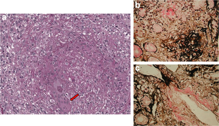
Fig. 6.
Renal biopsy from patient 20 of Table 1: a) The interlobular artery is extensively affected by a granulomatous necrotizing inflammatory process. The vessel wall is poorly recognizable due to the presence of wide areas of necrosis and leukocyte infiltration. Multinuclear giant cells can be seen in the infiltrate (arrow) (periodic acid Schiff, × 400). b) A small artery with fibrinoid necrosis of the wall and perivascular lympho-monocyte infiltration (methenamine silver, × 400). c) Fibrinoid necrosis of the wall of a small artery (methenamine silver, × 400)

