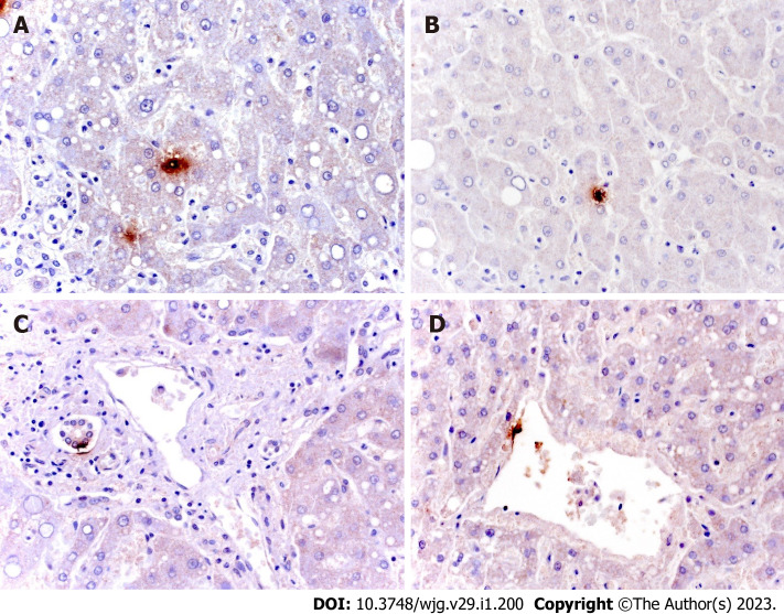
Figure 2.
Immunohistochemical staining for severe acute respiratory syndrome coronavirus 2 in liver tissue. A and B: Spot localization of virus in samples of initial hepatic necrosis and in Kupffer cells; C and D: Spot localization of isolated ductular and endothelial cells (mouse, GeneTex GTX632604, 1A9 clone, 1:100).

