
Figure 5.
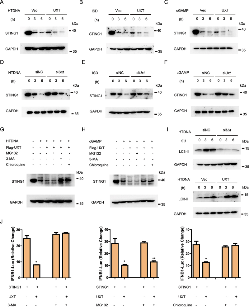
UXT promotes autophagic degradation of STING1. (A) MEFs transfected control vectors (Vec) or UXT expression plasmids (UXT) were stimulated with HTDNA (2 μg per well) for 0, 3, 6 h respectively. Then, Cell lysates were collected for western blot analysis of STING1 and GAPDH. (B) MEFs transfected control vectors (Vec) or UXT expression plasmids (UXT) were stimulated with ISD (2 μg per well) for 0, 3, 6 h respectively. Then, Cell lysates were collected for western blot analysis of STING1 and GAPDH. (C) MEFs transfected control vectors (Vec) or UXT expression plasmids (UXT) were stimulated with cGAMP (1 μg per well) for 0, 3, 6 h respectively. Then, Cell lysates were collected for western blot analysis of STING1 and GAPDH. (D) MEFs transfected negative control (NC) or Uxt siRNAs were stimulated with HTDNA (2 μg per well) for 0, 3, 6 h respectively. Then, Cell lysates were collected for western blot analysis of STING1 and GAPDH. (E) MEFs transfected negative control (NC) or Uxt siRNAs were stimulated with ISD (2 μg per well) for 0, 3, 6 h respectively. Then, Cell lysates were collected for western blot analysis of STING1 and GAPDH. (F) MEFs transfected negative control (NC) or Uxt siRNAs were stimulated with cGAMP (1 μg per well) for 0, 3, 6 h respectively. Then, Cell lysates were collected for western blot analysis of STING1 and GAPDH. (G) MEFs transfected control vectors (Vec) or UXT expression plasmids (UXT) were stimulated with HTDNA (2 μg per well) for 0, 3 h respectively, followed by the treatment of mock, MG132 (10 μM), chloroquine (50 μM), or 3-MA (10 mM). Then, Cell lysates were collected for western blot analysis of STING1 and GAPDH. (H) MEFs transfected control vectors (Vec) or UXT expression plasmids (UXT) were stimulated with cGAMP (1 μg per well) for 0, 3 h respectively, followed by the treatment of mock, MG132 (10 μM), chloroquine (50 μM), or 3-MA (10 mM). Then, Cell lysates were collected for western blot analysis of STING1 and GAPDH. (I) MEFs transfected negative control (NC) or Uxt siRNAs and control vectors (Vec) or UXT expression plasmids (UXT) were stimulated with HTDNA (2 μg per well) for 0, 3, 6 h respectively. Then, Cell lysates were collected for western blot analysis of LC3-II and GAPDH. (J) Luciferase activity in HEK293 cells transfected with an IFNB luciferase reporter, together with STING1 and an empty vector or UXT expressed plasmids, followed by the treatment of mock, MG132 (10 μM), chloroquine (50 μM), or 3-MA (10 mM). The firefly- and renilla luciferase signals were detected with Dual Glo® luciferase assay (Promega). Graphs show the mean ± SEM, and the data shown are representative of three independent experiments. *p < 0.05; **p < 0.01; ***p < 0.001 (Two-tailed t-test).