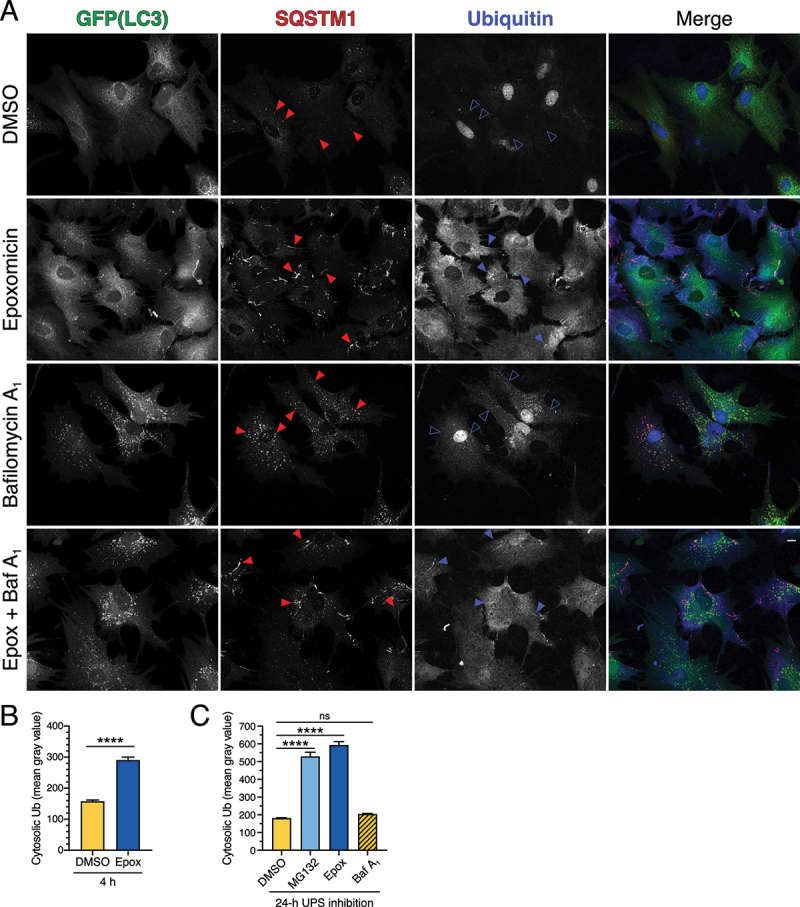
Figure 2.

SQSTM1 fibril-like structures that form in response to short-term UPS inhibition are partially positive for ubiquitin. (A, B) Immunostain analysis and corresponding quantification of primary astrocytes treated with 5 µM epoxomicin supplemented with 100 nM Baf A1 (or equivalent volume of DMSO as a solvent control) for 4 h. (A) Maximum projections of z-stacks of GFP-LC3 transgenic astrocytes immunostained for GFP, SQSTM1, and ubiquitin. Closed red arrowheads denote SQSTM1-positive puncta or fibril-like structures; closed blue arrowheads denote SQSTM1-positive fibril-like structures that are co-positive for ubiquitin; open blue arrowheads denote SQSTM1-positive puncta that are negative for ubiquitin. Bar: 10 µm. (B) Quantification of cytosolic ubiquitin signal intensity after 4 h of UPS inhibition (means ± SEM; unpaired t-test; n = 97–136 cells from 3 independent experiments; 6 DIV). (C) Quantification of cytosolic ubiquitin signal intensity after 24 h of UPS inhibition (0.5 µM MG132 or 10 nM epoxomicin) or after 4 h of 100 nM baf A1-treatment (means ± SEM; one-way ANOVA with Tukey’s post hoc test; n = 82–116 cells from 3 independent experiments; 3–8 DIV).
