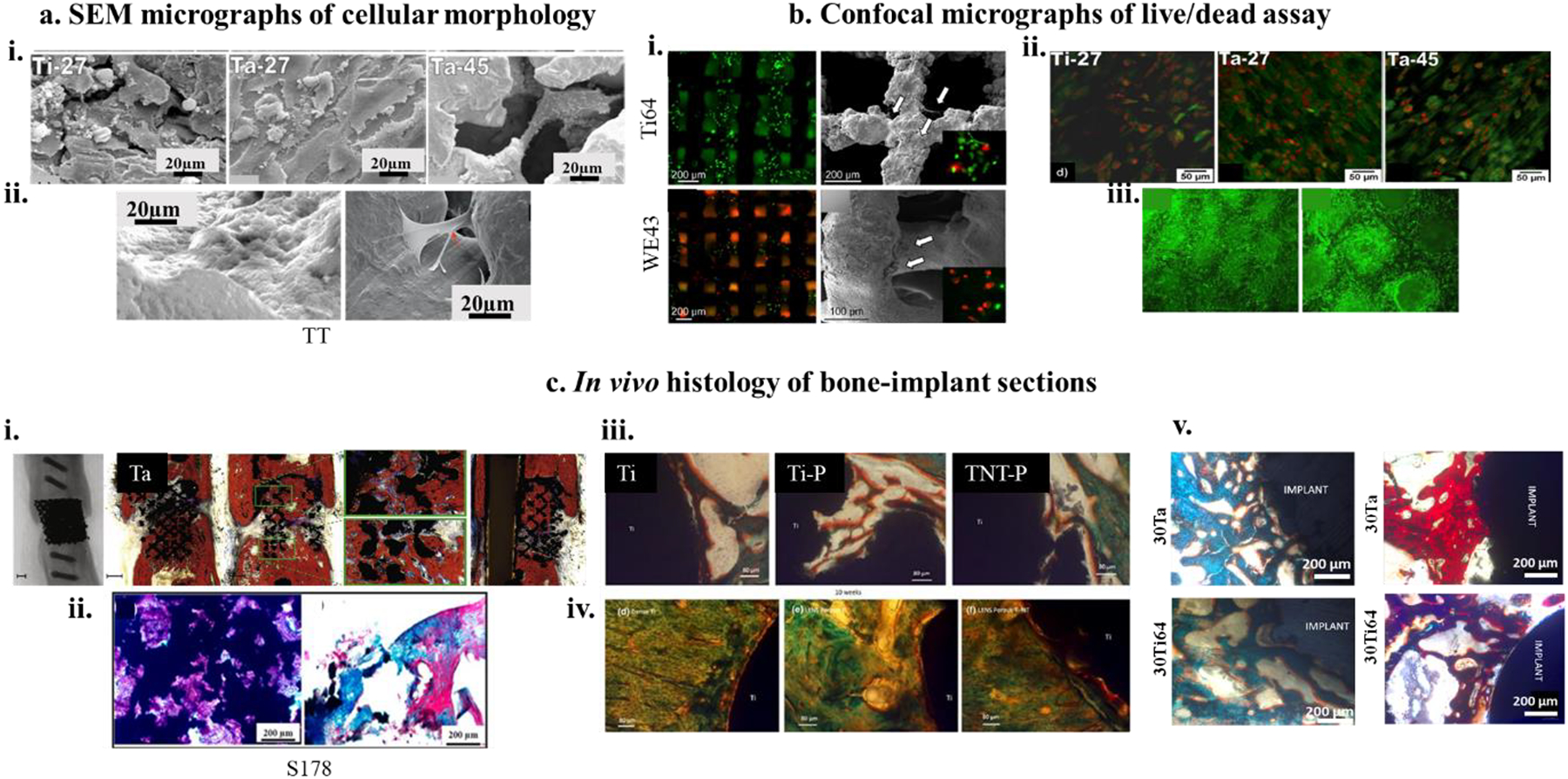
Figure 5. a. Cell-materials interaction as a function of cellular morphology investigation,

i) SEM micrographs of hFOB morphology on DED processed 27% porous Ti and Ta and 45% porous Ta at day 11, showing Ta-27 has the most optimized surface for cell-material interaction with cellular confluence as well as excellent proliferation4 ii) shows highly efficient focal adhesion of human adipose-derived stem cells (hADCs) on trabecular tantalum (TT) surface106. b) Fluorescence assay of dead and living cells on biocompatible metal surfaces. i) (Top left and bottom left) show fluorescence micrographs of Ti64 and Mg alloy (WE43) at 4h after seeding MG-63 osteoblast-like cells as compared to 24h fluorescence images (Top right and bottom right113) ii and iii) ALP protein expression for hFOB cells on 27% porous Ti and Ta and 45% porous Ta has shown through fluorescence micrographs. In contrast to SEM micrographs, Ta-27 did not show strong ALP expression at day 114. c) In vivo bone formation and osseointegration analyses, i) X-ray and histological images of open porous SLM-processed Ta revealing a fair amount of osseous growth for both the porous Ta specimens with almost 100% bridging of the defect85, ii) H&E (left) and Masson-Goldner (right) stained bone sections after 12 weeks of implantation of Ti64 implants with 178 μm pore size showing new bone formation around the porous metal implant,122 iii and iv) Photomicrograph showing the histology images after 4 weeks (top row) and 10 weeks (bottom row) for Ti, porous Ti (Ti-P) and porous Ti with nanotube surface (TNT-P) where signs of osteoid like new bone formation could be seen in orange/red color. Modified Masson Goldner’s trichrome staining method was used143,v) In vivo biological response from tantalum parts fabricated using direct energy deposition (DED) showing early-stage osseointegration at 5 weeks (left column) as a function of designed porosities and extended new bone formation at 12 weeks (right column)37.
