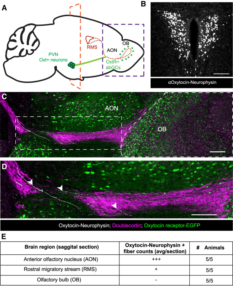
Figure 2.

Oxytocin+ fibers innervate the rostral migratory stream. (A) Schematic diagram of a tracing experiment. (PVN) Paraventricular nucleus of the hypothalamus, (RMS) rostral migratory stream, (AON) anterior olfactory nucleus, (OB) olfactory bulb. (B) Coronal slice of the PVN. The plane is denoted by the orange dashed line in F, showing Oxt-immunopositive neurons in the PVN. Scale bar, 100 µm. (C) Sagittal slice. The plane is denoted by the purple dashed line in A, showing Oxt-immunopositive fibers in white, Oxtr-EGFP-immunopositive cells in green, and doublecortin-immunopositive neurons in purple to label neurons within the RMS and OB. Scale bar, 200 µm. (D) Magnified view of the white box in C. White arrowheads show oxytocinergic fibers directly innervating the RMS. Scale bar, 200 µm. (E) Semiquantitative count of oxytocin−neurophysin+ fibers in the AON, RMS, and OB. (+++) 10+ fibers per section, (+) one to three fibers per section,(-) zero fibers per section.
