Figure 2.

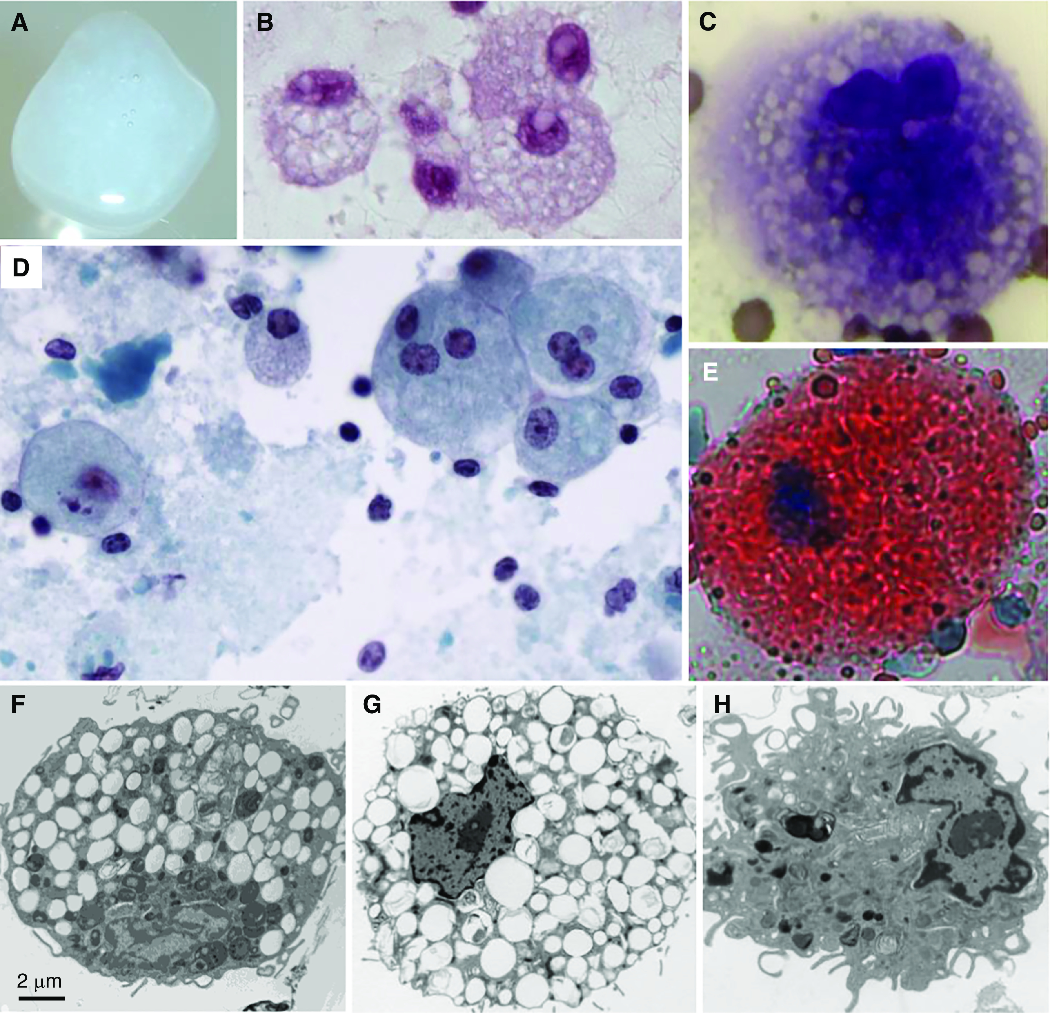
Sputum, sputum cytology, and alveolar macrophage ultrastructure in autoimmune pulmonary alveolar proteinosis (PAP). (A) Gross appearance of freshly expectorated sputum from a patient with autoimmune PAP. (B–E) Microscopic appearance of sputum cytology after staining with periodic acid–Schiff reagent (B); Diff-Quick, showing a foamy alveolar macrophage (C); Papanicolaou reagent (D); or oil red O, showing an oil red O–positive alveolar macrophage (E). (F) Alveolar macrophage obtained by BAL from an 18-year-old man with autoimmune PAP several months after lung transplantation performed as therapy for pulmonary fibrosis. Note the numerous, single membrane–delimited, intracytoplasmic lipid droplets. Scale bar, 2 μm. (G and H) Uranyl acetate staining. Alveolar macrophages obtained from a nonhuman primate passively immunized with highly purified, human GM-CSF (granulocyte/macrophage colony–stimulating factor) autoantibodies derived from patients with autoimmune PAP, showing intracytoplasmic lipid droplets (G), or a saline-injected primate, showing a normal macrophage appearance (H).
