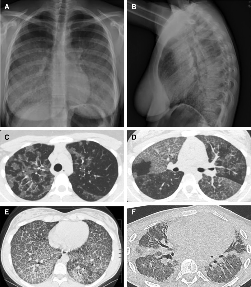
Figure 3.
Appearance of the chest radiograph and chest computed tomography scan in autoimmune pulmonary alveolar proteinosis (PAP). (A and B) Posterior–anterior (A) and lateral (B) chest radiographs of a 19-year-old woman with autoimmune PAP showing diffuse ground-glass opacification of the lung parenchyma. (C–F) Representative images from computed tomography of the chest showing the diversity of radiographic findings in autoimmune PAP in a 45-year-old man (C), a 58-year-old woman (D), a 15-year-old girl (E), and an 18-year-old man (F). (C) Image showing ground-glass opacification involving some but not all secondary lobules resulting in a distinctive “geographic” pattern. Also, note the disproportionate involvement of the left and right lung parenchyma. (D) Image showing a distinctive pattern of interlobular septal thickening superimposed on ground-glass opacification, often referred to as “crazy paving.” Also, note the sharply demarcated differences in the degree of involvement between adjacent lung lobes. (E) Image revealing a homogeneous pattern of crazy paving throughout all regions of the lung parenchyma. (F) Image revealing extensive pulmonary fibrosis with parenchymal distortion from traction bronchiectasis. This patient underwent bilateral lung transplantation for pulmonary fibrosis and respiratory failure shortly after this image was obtained.

