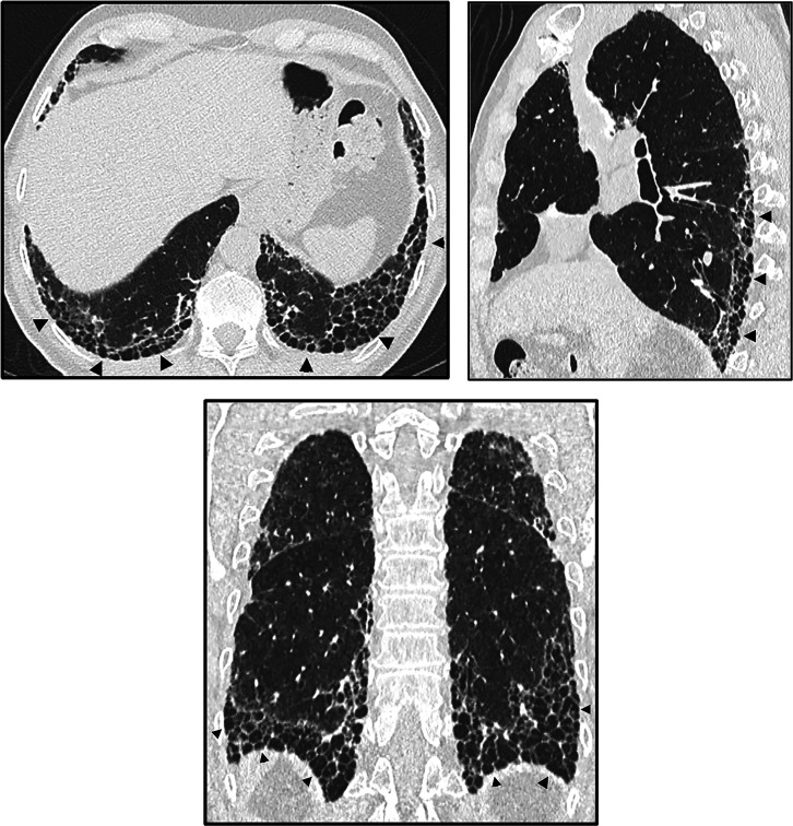
Figure 2.
Honeycombing. Axial, sagittal, and coronal computed tomography images show subpleural-predominant, lower lung–predominant reticular abnormality with honeycombing (arrowheads). Honeycombing is defined by clustered, thick-walled, cystic spaces of similar diameters, measuring between 3 and 10 mm but up to 2.5 cm in size. The size and number of cysts often increase as the disease progresses. Often described in the literature as being layered, a single layer of subpleural cysts is also a manifestation of honeycombing. Honeycombing is an essential computed tomography criterion for typical (“definite”) usual interstitial pneumonia–idiopathic pulmonary fibrosis pattern when seen with a basal and peripheral predominance. In this pattern, honeycombing is usually associated with traction bronchiolectasis and a varying degree of ground-glass attenuation.

