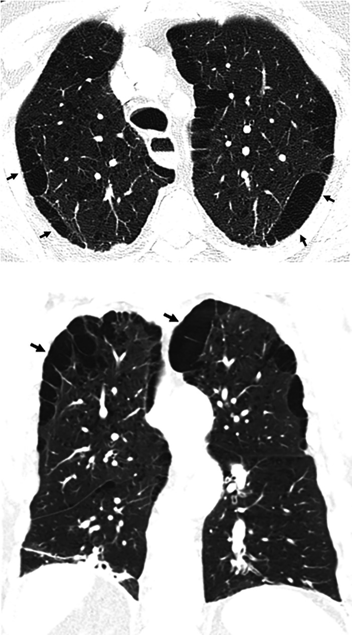
Figure 3.

Paraseptal emphysema. Axial and coronal computed tomography images show relatively large subpleural cysts of paraseptal emphysema (arrows), mainly in the upper lobes. Centrilobular emphysema is also present. The subpleural cysts of paraseptal emphysema usually occur in a single layer and are larger than honeycomb cysts (typically >1 cm); they are not associated with other features of fibrosis such as reticular abnormality or traction bronchiectasis.
