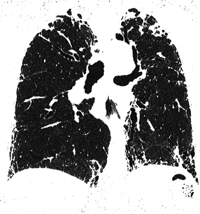
Figure 7.

Combined pleuroparenchymal fibroelastosis and usual interstitial pneumonia patterns. Coronal computed tomography image shows dense subpleural fibrosis at the lung apices with traction bronchiectasis and upper lobe volume loss. There is subpleural reticular abnormality and honeycombing in both lower lobes.
