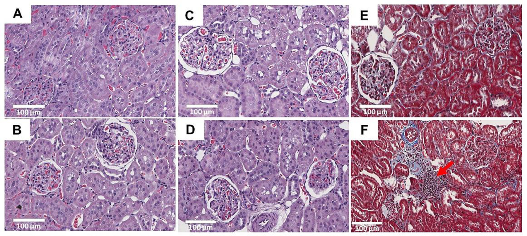
Figure 5.

H&E and trichrome staining of kidney sections (20x) in twelve weeks of experimental animals. No significant changes were observed between control (A) and H-MSG group (B, C, D). No significant differences in interstitial fibrosis was seen in between control (E) and H-MSG group (F) animals. Marked infiltration of mononuclear inflammatory cells (red arrow) in the interstitial space of renal tubules were observed in H-MSG group compared to control.
