. 2023 Jan 19;12:e82843. doi: 10.7554/eLife.82843

Figure 6. Model of hypoxia-induced SM truncation.

(A, B) Hypoxic conditions stabilize the E3 ubiquitin ligase MARCHF6 and inhibit SM-catalyzed conversion of squalene to monooxidosqualene, leading to squalene accumulation. (C) Increased MARCHF6 activity promotes ubiquitination of SM and its targeting to the proteasome. (D) Squalene impedes the complete proteasomal degradation of SM via a mechanism involving the SM-N100 domain, (E) yielding the constitutively active trunSM. During transient or low-level hypoxia, trunSM activity may facilitate continued cholesterol synthesis to compensate for the oxygen shortfall. During long-term or severe hypoxia, trunSM activity may reduce squalene-induced toxicity and promote downstream synthesis of lanosterol, which suppresses an early step of the cholesterol synthesis pathway. During pathophysiological hypoxia, cholesterol synthesis enabled by trunSM may contribute to oncogenic cell growth and survival.

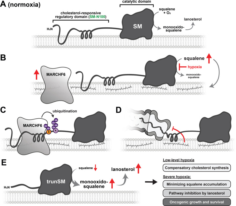
Figure 6.

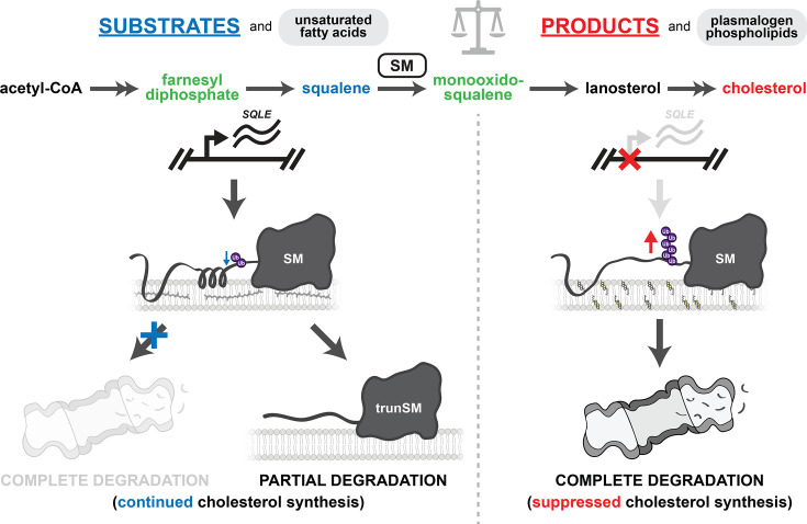
Figure 6—figure supplement 1. Summary of metabolic regulation of SM.

Figure 6—figure supplement 1.

Squalene (blue), other farnesyl-containing cholesterol synthesis intermediates (green), and unsaturated fatty acids stabilize SM by impeding its ubiquitination and complete proteasomal degradation. When SM reaches the proteasome, squalene and farnesyl-containing intermediates can also promote its partial degradation to yield the constitutively active trunSM. In both cases, SM activity is preserved and continued cholesterol synthesis is favored. Excess cholesterol suppresses SQLE gene expression and accelerates the ubiquitination and complete proteasomal degradation of SM. Plasmalogen phospholipids also promote SM ubiquitination and degradation. This suppresses SM activity and downstream cholesterol synthesis. Balance of these feedforward and feedback loops ensures cholesterol synthesis is tightly coupled with supply and demand.