
FIGURE 1.
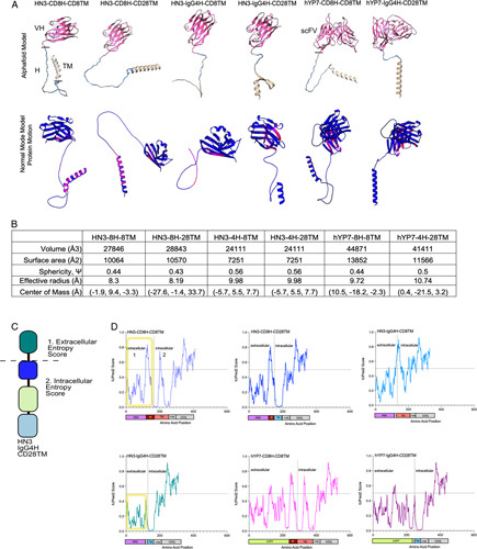
Structural modeling and entropy analysis reveals that the IgG4H and CD28TM display limited flexibility and low entropy. (A) Alphafold models and normal mode analysis of protein movement. The VH or scFV are coded in pink, the hinge (H) is in blue and the transmembrane (TM) is in tan. Residues with higher movement are listed in red, while restricted residues are in blue. (B) Physical characteristics of each CAR based on the models. Cell constructs are abbreviated as 8H-8TM (CD8H, CD8TM), 4H-28TM (IgG4H, CD28TM), 4H-8TM (IgG4H, CD8TM). (C) Diagram of extracellular and intracellular domains in the CAR T cell. The 4 main components include the targeting element, hinge, transmembrane, and t cell signaling portions. The intracellular and extracellular entropies are measured in 1 and 2. (D) Entropy scores using IUPRED2 algorithm. A higher score corresponds to higher entropy, while a lower score denotes lower entropy. CAR, chimeric antigen receptor.