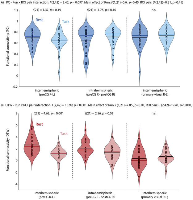
Fig. 3.
FC between cortical motor regions (pre- and post-central gyrus from the Harvard-Oxford atlas) during a resting state and a task fMRI in the same participants. A) Interhemispheric FC (preCG R-L) was reduced during the task compared to rest for DTW estimates of FC, as was expected given frequent button presses with the left index and middle finger during task fMRI. It did not differ for PC estimates of FC. Results of post-hoc pairwise paired-samples t-tests are reported for each comparison. B) Interhemispheric connectivity between left and right primary visual cortex was also estimated as a control and did not differ between rest and task fMRI for PC or DTW estimates of FC. Note, that for DTW estimates a value of 0 reflects average FC while positive values indicate above average time series similarity.

