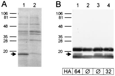
FIG. 6.
Coomassie blue-stained SDS-PAGE (A) and immunoblot analysis using anti-SfpA antiserum (B) of thermoeluted fimbrial extracts taken from recombinant E. coli strains HB101/pSFO157-E11 (lane 1), HB101/pBluescript II KS (vector control) (lane 2), HB101/pSFO157-ESn8 (sfpG mutant) (lane 3), and pSFO157-ESn8::sfpGApa (lane 4). Positions and sizes of marker proteins (in kilodaltons) are given on the left. The arrows indicate the SfpA protein as identified by amino acid sequencing. Titers of mannose-resistant hemagglutination (HA) of the strains are given below the immunoblot. The bands strongly reacting with anti-SfpA antiserum above and below the SfpA protein and not visible in Coomassie blue-stained SDS-PAGE are suspected to represent nonproteinaceous components of the bacterial cell envelope present in the SfpA preparation used for immunization. These bands were not detected using rabbit normal serum, nor were they visible in silver-stained gels.

