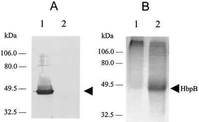
FIG. 5.
Characterization of recombinant fusion HbpB protein. (A) Total protein samples were prepared from the cells indicated and then used in Western blotting as described in the legend for Fig. 4A. Lanes: 1, protein from E. coli BL21(DE3) with pSH9; 2, protein from E. coli BL21(DE3) with pRsetA vector alone. (B) The indicated protein samples were subjected to LDS-PAGE and then reacted with TMBZ. Lanes: 1, 10 μg of soluble protein from E. coli BL21(DE3) with pRsetA vector alone; 2, 10 μg of soluble protein from E. coli BL21(DE3) with pSH9. Both samples were preincubated with hemin before electrophoresis. The 46-kDa HbpB fusion protein is marked by an arrow.

