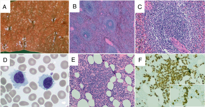
Fig. 3.
Splenic marginal zone lymphoma. On gross examination, the splenic parenchyma shows increased and expanded white pulp nodules (A). On H&E sections, the splenic parenchyma shows increased numbers of white pulp nodules (B), which are typically composed of central cores containing small lymphocytes with scant cytoplasm and a peripheral zone of marginal zone cells with more abundant cytoplasm (C, H&E). In the peripheral blood, the neoplastic cells are small, often with a somewhat plasmacytoid appearance, and, in some cases, cytoplasmic projections may be seen (D, Wright’s stain). In trephine biopsy samples, interstitial lymphoid aggregates are present (E, H&E) with numerous small B-cells seen within the aggregate (F, CD20). Often, at least focal areas will display an intrasinusoidal growth pattern of small B-cells (inset, F)

