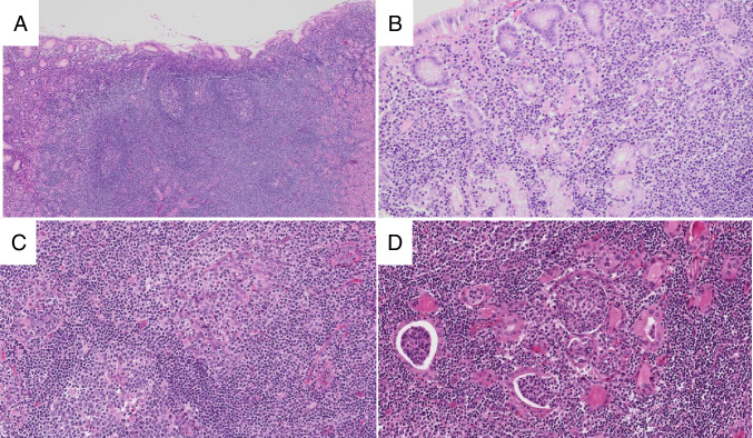
Fig. 4.
Extranodal marginal zone lymphoma. In gastric MALT lymphoma, the mucosa contains a dense infiltrate of small lymphocytes and germinal centers (A, H&E). The small lymphocytes form desctructive lymphoepithelial lesions (B, H&E). In this salivary gland MALT lymphoma (C, H&E), the lymphoid proliferation contains germinal centers (lower left) and prominent lymphoepithelial lesions (upper right). In thyroid MALT lymphomas (D, H&E), lymphoepithelial lesions consist of small lymphocytes within glandular lumens (so-called MALT balls)

