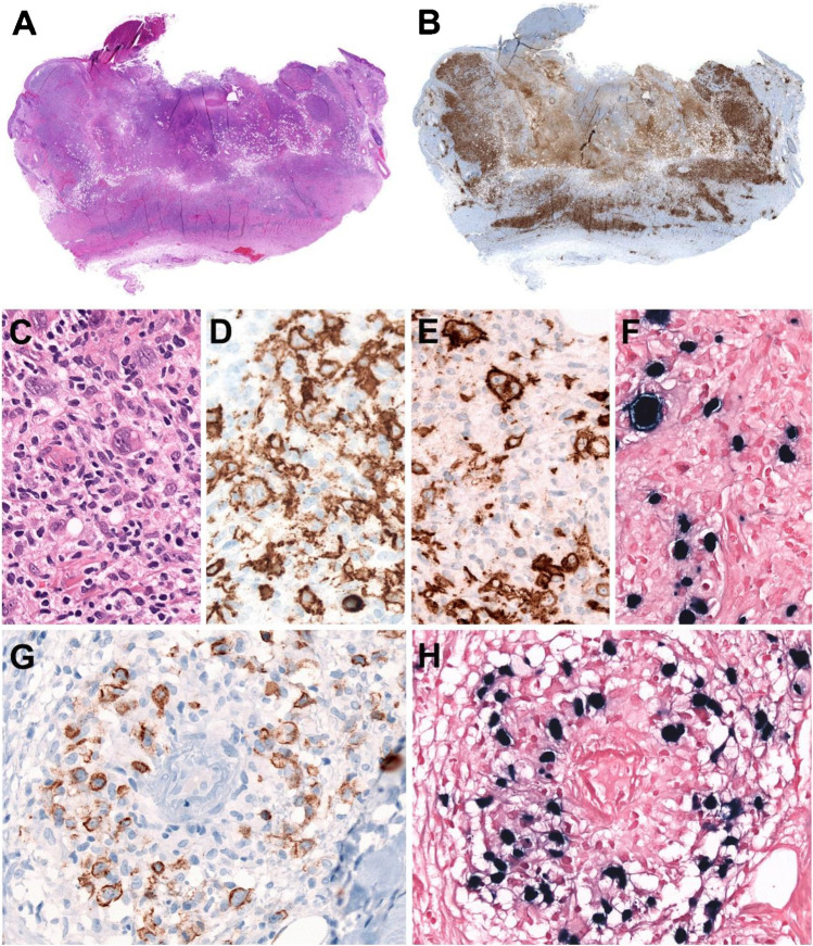
Fig. 1.
EBV-positive mucocutaneous ulcer. A solitary skin lesion in the forehead of a 70-year-old man without known immunosuppression. A Panoramic view of a well-circumscribed ulcer (snap-shot from scanned slide). B CD3 stain demonstrates abundant reactive T cells that form a rim at the base of the ulcer (snap-shot from scanned slide). C Higher magnification showing Hodgkin and Reed–Sternberg (HRS)-like cells in a polymorphic background. D CD20 stain is positive in the large HRS-like cells and in medium and small B cells. E CD30 stain is strongly positive in the HRS-like cells. F EBER in situ hybridization is positive in the HRS-like cells. Note the wide range of cells positive for EBER. G LMP1 is positive in B cells surrounding a blood vessel (angiocentric lesion). H The LMP1-positive cells are also positive for EBER. (C–H, original magnification, 400×)

