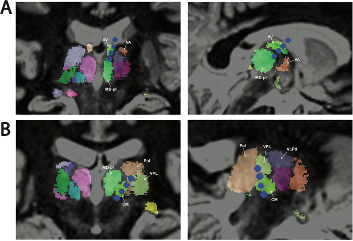
Figure 2.

Targeted thalamic nuclei delineated in atlas space. (A) Anterior nucleus of the thalamus (ANT) was targeted with a frontal approach. The mammillothalamic tract is targeted surgically. Two in‐line views shown. (B) Centromedian nucleus of the thalamus (CMT) was targeted with a posterolateral approach using the T2 white‐matter‐null MRI sequence. Two in‐line views shown. Coordinates are as follows: reference point PC, target CM coordinates: lateral left 9.07 mm, anterior 1.55 mm, inferior 1.39 mm; direction lateral left 34.26 degrees, posterior 13.99 degrees, and trajectory length 71.18 mm. AC‐PC distance 23.79 mm. Electrode contacts are in blue. Relevant nuclei are labeled. Nuclei were derived using the THOMAS atlas 17 and LeGui software (see Methods section for details). 18 AV: anterior ventral nucleus, CM: centromedian nucleus, MD‐pf: mediodorsal‐parafascicular nucleus, Pul: pulvinar nucleus., VA: ventral anterior nucleus, VPL: ventral posterior lateral nucleus, VLPd: ventral lateral posterior dorsal group.
