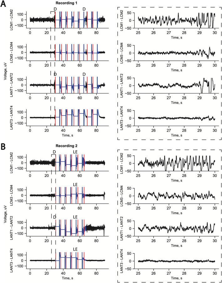
Figure 3.

Example long‐episode ECogs from patient's RNS data. Ictal activity is consistently recorded from CMT channel 1–2 before ANT channel 1–2 in both examples. Right side insets show blown up timeline. In both examples, the seizure is aborted with stimulation across all four channels. (A) Example seizure detected first with ictal discharge recorded across the distal contact pair LCM1‐LCM2 (in CMT proper) followed by ictal activity in ANT1‐ANT2 (in MD and ANT) at 29 s and ictal activity recorded in LCM3‐LCM4 (within pulvinar) at 29.5 s. ANT3‐ANT4 (in the ventricle) is electrically quiet as expected. (B) Again, ictal activity is seen in the distal contact pair LCM1‐LCM2 (in CMT proper) followed by ictal activity in ANT1‐ANT2 (in MD and ANT) at 29 s. CMT3‐CMT4 shows ictal discharges. ANT3‐ANT4 is electrically quiet. D: detection, LE: long episode, red marks: stimulation treatment delivery.
