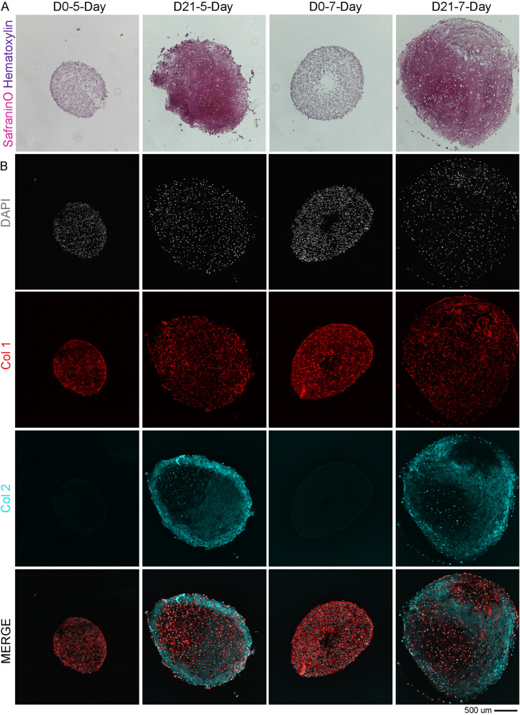
Fig. 4.
Minimum expansion time frame to obtain chondrogenic hADSCs from the isolation phase. A Representative brightfield images of cryosections from pellet culture stained with SafraninO (in pink) and Haematoxylin (in purple) to identify cell’s nuclei. B Representative confocal images of cryosections from pellet immunostained with Phalloidin-RFP (Actin, in red), Collagen type 2 (Col 2, in cyan) and counterstained to detect cells nuclei (DAPI, in white). Superimposed channels are shown in the last row of panels (MERGE). For all the stainings, the cryosections were obtained from cells pelleted after 5 and 7 days of non-passaged proliferation, pushed into 3 weeks of chondrogenic differentiation. The calculated Collagen type 2 intensity from day 0 to day 21 in reported as Fold Increase (F.I.) in the day 21 panels of their corresponding groups

