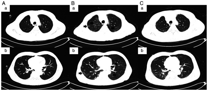
Figure 2.
CT images of the patient. (A-a and A-b) CT images showing interstitial changes in both lungs at admission following 6 months of cessation of immunotherapy (September 2021). (B-a and B-b) CT revealed interstitial changes and multiple ground-glass opacities (indicated by arrows in the images) in both lungs 10 days after anti-infection treatment (September 2021). (C-a and C-b) CT showing progressive improvement of immune checkpoint inhibitor-related pneumonitis after corticosteroid administration without recurrence (December 2021). A-a and A-b, B-a and B-b, and C-a and C-b are two representative examples shown for each case.

