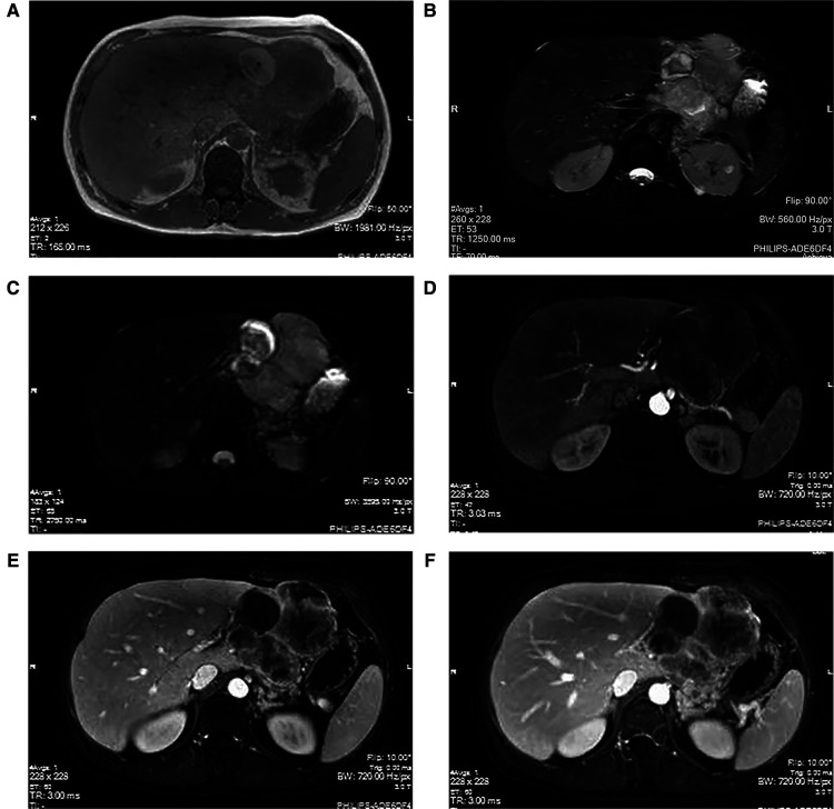
Figure 1.
MRI manifestations of hepatic sarcomatoid carcinoma. The magnetic resonance imaging (plain scan + enhanced + functional imaging) shows abnormal signals of irregular mass in the liver, with a size of approximately 9.5 × 8.3 cm. The T1WI (A) shows a weak signal; however, the T2WI (B) and the DWI (C) show strong signals. The arterial phase (D) indicates slightly uneven enhancement at the edge of the lesion, with continuous uneven enhancement in the portal phase (E) and delayed phase (F).

