. 2022 Dec 20;11(1):A0105. doi: 10.5702/massspectrometry.A0105

Fig. 3. (A) Brain tissue section with melanoma administered with BPA. MALDI-MSI was performed in the half area enclosed by square. The spotted BPA standard (upper: 100 pmol, lower: 20 pmol) outside brain section were used to confirm the detection of BPA (B) tumor area designated as a region of interest (ROI).
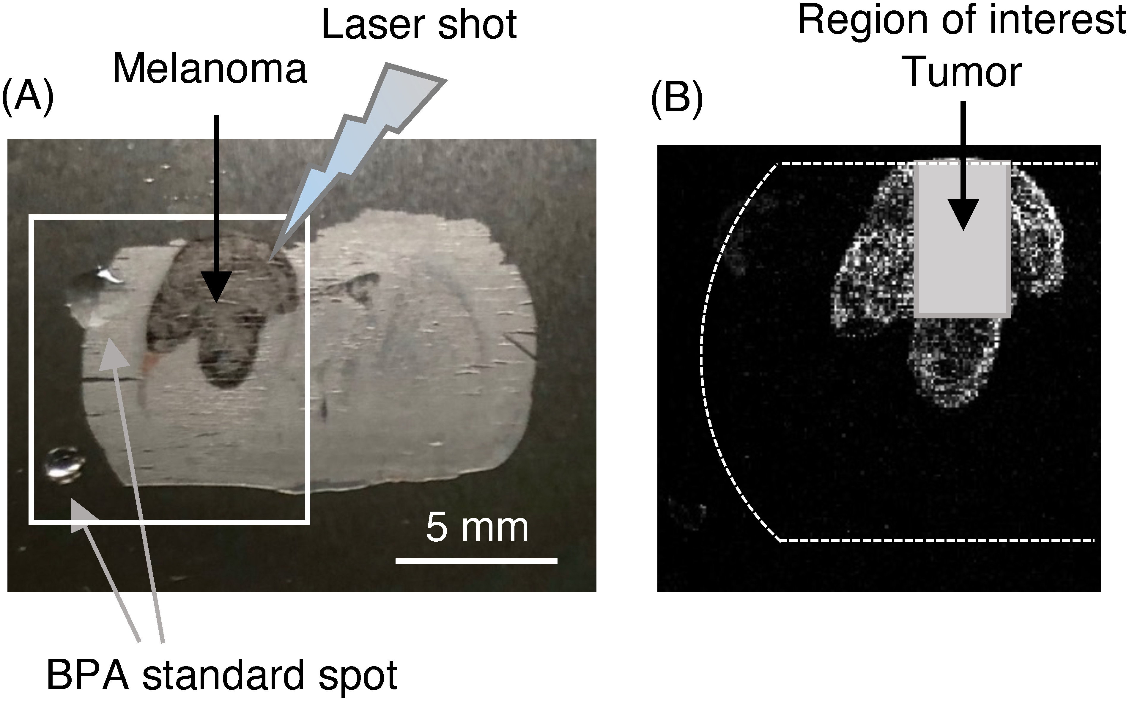
Fig. 3. (A) Brain tissue section with melanoma administered with BPA. MALDI-MSI was performed in the half area enclosed by square. The spotted BPA standard (upper: 100 pmol, lower: 20 pmol) outside brain section were used to confirm the detection of BPA (B) tumor area designated as a region of interest (ROI).