Figure 6.
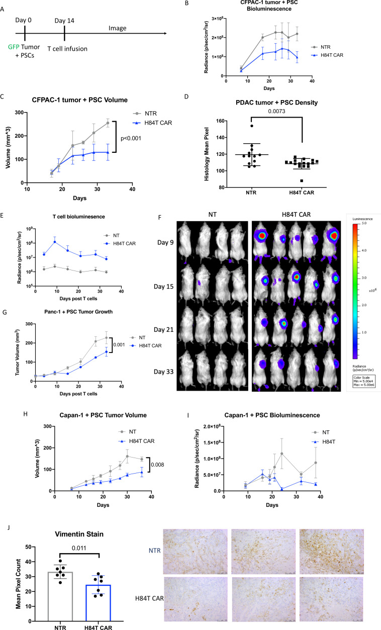
In vivo antitumor and anti-stromal activity of H84T CAR-T cells against three different PDACs. (A) NSG MHC KO mice were engrafted subcutaneously with 1×10⁶ GFP-FfLuc labeled CFPAC-1 tumor cells and 1×10⁶ PSCs. Tumors were allowed to establish for 2 weeks and then 1×10⁶ NT or H84T CAR-T cells were intravenously delivered. (B) Tumor signal was quantified by bioluminescence signaling through IVIS imaging. N=4 mice per group. (C) Tumor volume was measured by the caliper and volume was calculated overtime. P value<0.001 determined by simple linear regression. (D) Residual tumors were resected on Day 36 post T-cell infusion and processed for H&E staining. Mean pixel density of H&E staining was quantified by ImageJ. Four sections per each tumor were quantified and graphed. N=3 tumors/NTR group and N=4 tumors/H84T group. P value determined by unpaired student’s t-test. (E, F) NSG MHC KO mice were engrafted subcutaneously with 2×10⁶ Panc-1 tumor cells and 2×10⁶ PSCs. Tumors were allowed to establish for 2 weeks and then 1×10⁶ NT or H84T CAR-T cells labeled with GFP-FfLuc were delivered intravenously. T-cell signal was quantified by bioluminescence signaling through IVIS imaging (F) bioluminesence images. (G) Tumor volume was measured by the caliper and volume was calculated overtime. N=4–5 mice per group. (H, I) NSG MHC KO mice were engrafted subcutaneously with 3×10⁶ GFP-FfLuc labeled Capan-1 tumor cells and 3×10⁶ PSCs. Tumors were allowed to establish for 1 week and then 1×10⁶ NT or H84T CAR-T cells were intravenously delivered. Tumor volume (H) was quantified by caliper measurement and tumor signal (I) was quantified by bioluminescence signaling through IVIS imaging. N=4 mice per group. (J) Residual CFPAC-1+PSC tumors from mice treated with either NT or H84T CAR T cells were stained for anti-human vimentin to detect stroma cells. Vimentin pixel count was calculated by ImageJ analysis and representative IHC images are shown for three mice in each group. CAR, chimeric antigen receptor; PDAC, FfLuc, firefly luciferase; PDAC, pancreatic ductal adenocarcinoma; PSC, pancreatic stellate cells; NT, non-transduced; IHC, immunohistochemistry; GFP, green fluorescent protein.

