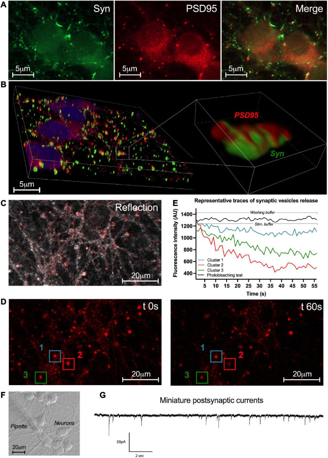
FIGURE 6.
Synaptic network activity of enteric neurons. (A) Double immunostaining of enteric neurons at D7 with the pre-synaptic marker synapsin I (Syn; green) and the post-synaptic marker PSD95 (red) (n = 3 culture wells from 3 independent rat embryos). (B) 3D reconstructed confocal microscopy images of cultured neurons immunolabeleled for synapsin 1 (green) and PSD95 (red) with a higher magnification of the boxed region. (C) Representative image of the enteric neuron culture in reflection microscopy. (D) Fluorescence images of a field containing clusters measured at t 0s and t 60s after KCl stimulation. (E) Plot of FM1-43 fluorescence intensity analyzed from a representative cluster recorded in washing buffer and 3 representative clusters recorded in stimulation buffer of the enteric neuron culture at D7 (154 clusters analyzed from n = 3 culture wells from 3 independent rat embryos). (F) Representative image of enteric neurons in light microscopy and the patch clamp pipette used for the whole-cell configuration. (G) Representative trace of miniature post-synaptic currents (mPSCs) recorded from enteric neurons at D7 (10 neurons analyzed from n = 5 culture wells from 5 independent rat embryos).

