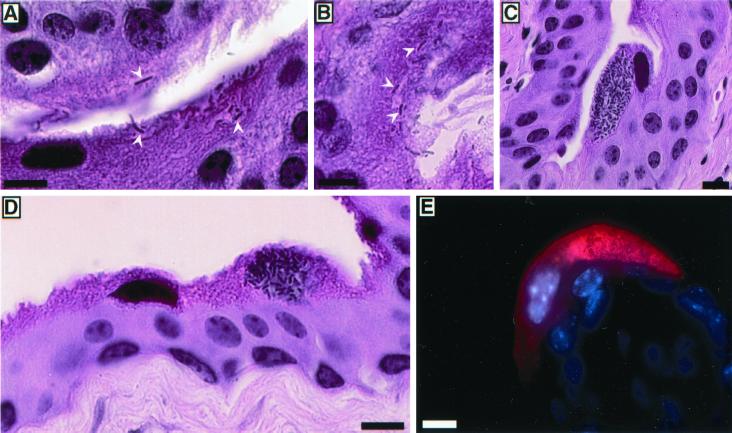
FIG. 2.
Replication of UPEC within superficial bladder cells. (A and B).Two hours after infection of C57BL/6 mice with UTI89, bacteria were detected in hematoxylin-and-eosin-stained sections entering or already within the superficial epithelial cells lining the lumenal surface of the bladder (arrowheads). (C to E) By 6 h after inoculation, large foci of intracellular E. coli were apparent within many of the superficial bladder cells. (E) Bacteria (red) were stained using an anti-E. coli primary antibody and Cy3-labeled secondary antibody. Host cell nuclei were visualized using Hoechst dye. Bars, 10 μm.

