
Figure 6.
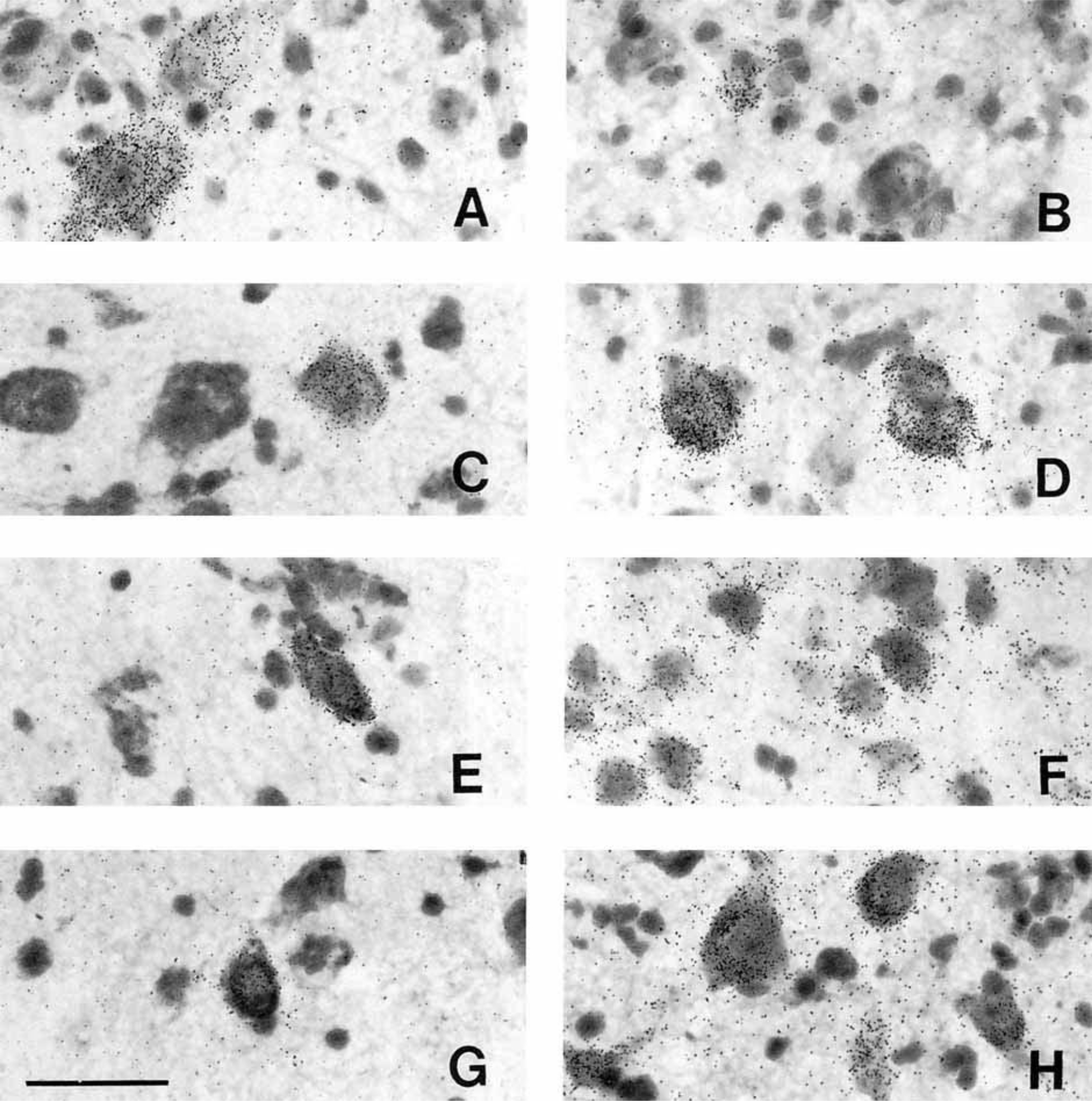
Photomicrographs of PDYN neurons in the human hypothalamus. A: Moderately and lightly labeled magnocellular neurons of the caudal paraventricular nucleus. B: Lightly labeled, small, peripherally located neuron in the dorsolateral supraoptic nucleus. C: Typical PDYN neuron of the tuberomammillary nucleus. D: Large, heavily labeled neurons of the posterior hypothalamic nucleus (coronal view). E: One of the few PDYN neurons of the retrochiasmatic area. F: PDYN neurons of the premammillary nucleus. G: Lightly labeled neuron of the ventromedial nucleus. H: PDYN neurons of the dorsomedial nucleus. Scale bar = 50 microns.