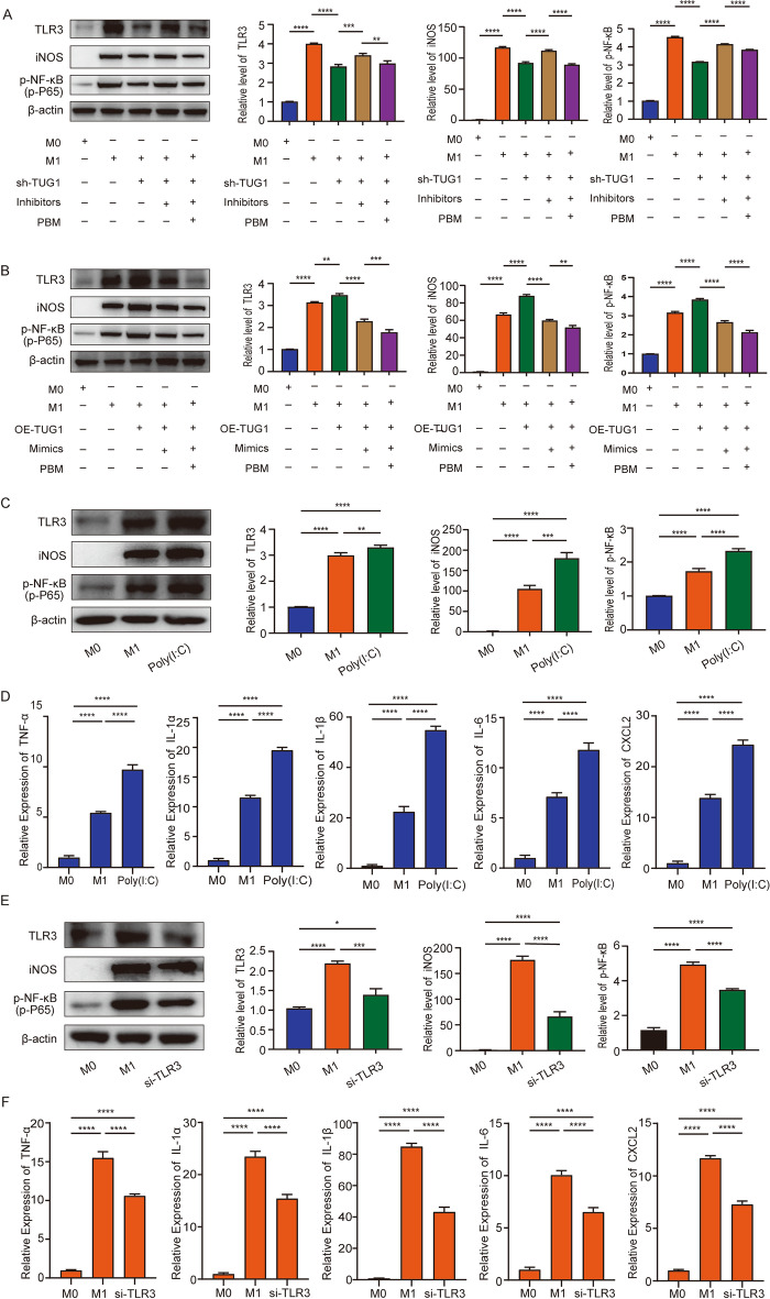
Fig. 6.
TUG1-miR-1192/TLR3 axis regulates macrophage polarization and inflammation. A Western blotting was used to detect the expression level of TLR3, iNOS, and p-NF-κB (p-p65), which was rescued by shTUG1 and miR-1192 inhibitor treatment. B Western blotting was used to detect the expression level of TLR3, iNOS, and p-NF-κB, which was rescued by OE-TUG1 and miR-1192 mimics treatment. C, D After overexpression of TLR3, western blotting was used to detect the expression of TLR3, iNOS, and p-NF-κB. RT-PCR detected the expression of TNF-α, IL-1α, IL-1β, IL-6, and CXCL2. E, F After knockdown of TLR3, western blotting was used to detect the expression of TLR3, iNOS, and p-NF-κB. RT-PCR detected the expression of TNF-α, IL-1α, IL-1β, IL-6, and CXCL2. n = 3 per group. *P < 0.05, **P < 0.01, ***P < 0. 001, ****P < 0.0001

