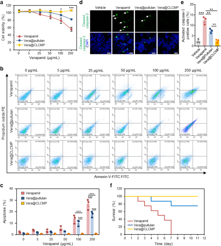
Fig. 3.
In vitro and in vivo toxicity of Vera@pullulan and Vera@CLCMP patches. a-c HepG2 cells were treated with the indicated concentration of verapamil dissolved in PBS, Vera@pullulan, and Vera@CLCMP patches, respectively, for 24 hrs. A Cell viability was measured by the WST-8 assay. B HepG2 cells were stained with annexin V-FITC and PI and then analyzed for apoptosis by flow cytometry. The percentage of apoptotic HepG2 cells are shown in (C). D Immunofluorescence staining for cleaved caspase-3 (green) in HepG2 cells treated with 250 μg/mL verapamil, 250 μg/mL Vera@pullulan, 250 μg/mL Vera@CLCMP, or PBS (vehicle) for 24 hrs. Nuclei were stained with DAPI (blue). White arrows show cleaved caspase-3 positive cells. Scale bar, 40 μm. E Caspase-3 activity was quantified. F Kaplan–Meier survival curve of mice treated daily with verapamil (50 mg/kg intraperitoneal [i.p.]), 50 mg/kg Vera@pullulan, and 50 mg/kg Vera@CLCMP patches, respectively, on day 0 (n = 6–14). Data is shown as mean ± SEM. *p < 0.05; ***p < 0.001 (One-way ANOVA, followed by Tukey’s test)

