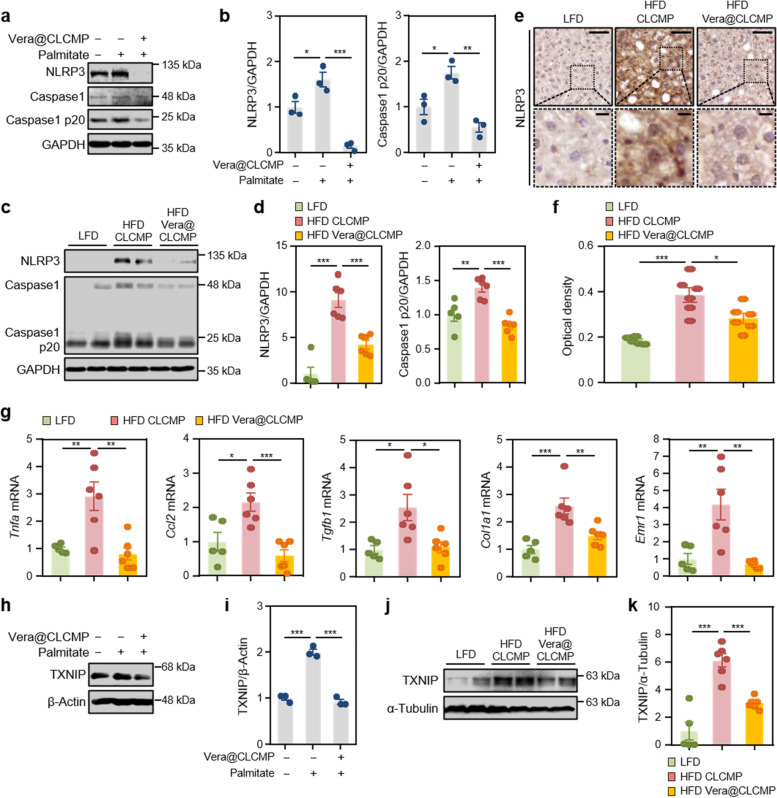
Fig. 9.
Vera@CLCMP patches ameliorate NLRP3-inflammasome activation by inhibiting the expression of TXNIP. A, B, H, I HepG2 cells were treated with 100 μM palmitate or BSA (vehicle) for 24 h in the presence or absence of a release medium incubated with 38.35 μg/mL Vera@CLCMP at 37 °C for 6 days. Cell lysates were immunoblotted with anti-NLRP3 and anti-caspase-1 (A) or anti-TXNIP (H) antibodies. GAPDH or α-tubulin served as a loading control. B, I Band intensities were quantified and normalized to control band intensities. C-G, J, K C57BL/6 male mice kept on HFD for 9 weeks had Vera@CLCMP or CLCMP patches applied on their dorsal skin. LFD-fed mice of the same age were used as a negative control. C Liver tissue lysates were immunoblotted with anti-NLRP3 and anti-caspase-1 antibodies. GAPDH served as a loading control. D Band intensities were quantified and normalized to control band intensities. E Immunohistochemical staining for NLRP3 in liver tissues from mice in each group indicated. Boxed areas are magnified in the bottom panels. Scale bars, 50 μm; 10 μm (insets). F Optical density of NLRP3 immunoreactivity. G qRT-PCR analysis of Tnfa, Ccl2, Tgfb1, Col1a1, and Emr1 mRNA levels in liver tissues from mice in each group indicated. J Liver tissue lysates were immunoblotted with anti-TXNIP antibody. α-tubulin served as a loading control. K Band intensities were quantified and normalized to control band intensities. Data is shown as mean ± SEM. *p < 0.05; **p < 0.01; ***p < 0.001 (One-way ANOVA, followed by Tukey’s test)

