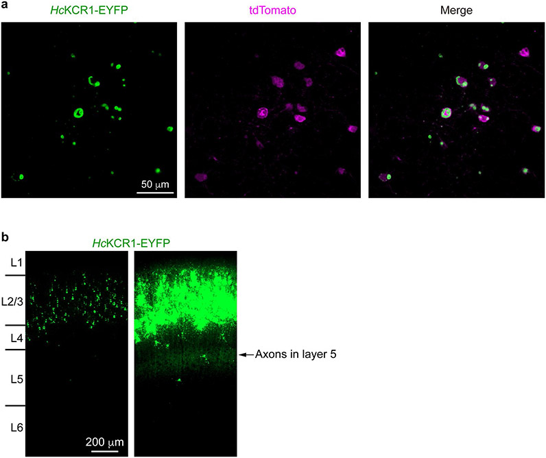
Extended Data Fig. 8 ∣. HcKCR1 expression in mouse cortical neurons.
a, Fluorescence images showing HcKCR1-EYFP (green) and tdTomato (magenta) expression in layer 2/3 pyramidal neurons. HcKCR1-EYFP is expressed at high levels and forms some intracellular aggregates, as do many other wild-type ChRs. HcKCR2-EYFP shows the same degree of aggregation. Membrane targeting of both KCRs is confirmed by robust photocurrents (Fig. 5b and Extended Data Fig 9.). b, The fluorescence image of HcKCR1-EYFP from Fig. 5a (left panel) was overexposed to visualize the presence of HcKCR1-EYFP in the dendrites and axons (right panel). Note, the axons of layer 2/3 pyramidal neurons ramify in layer 5. Similar results were observed in 14 slices from 2 male and 2 female mice at the age of 3–4 weeks.

