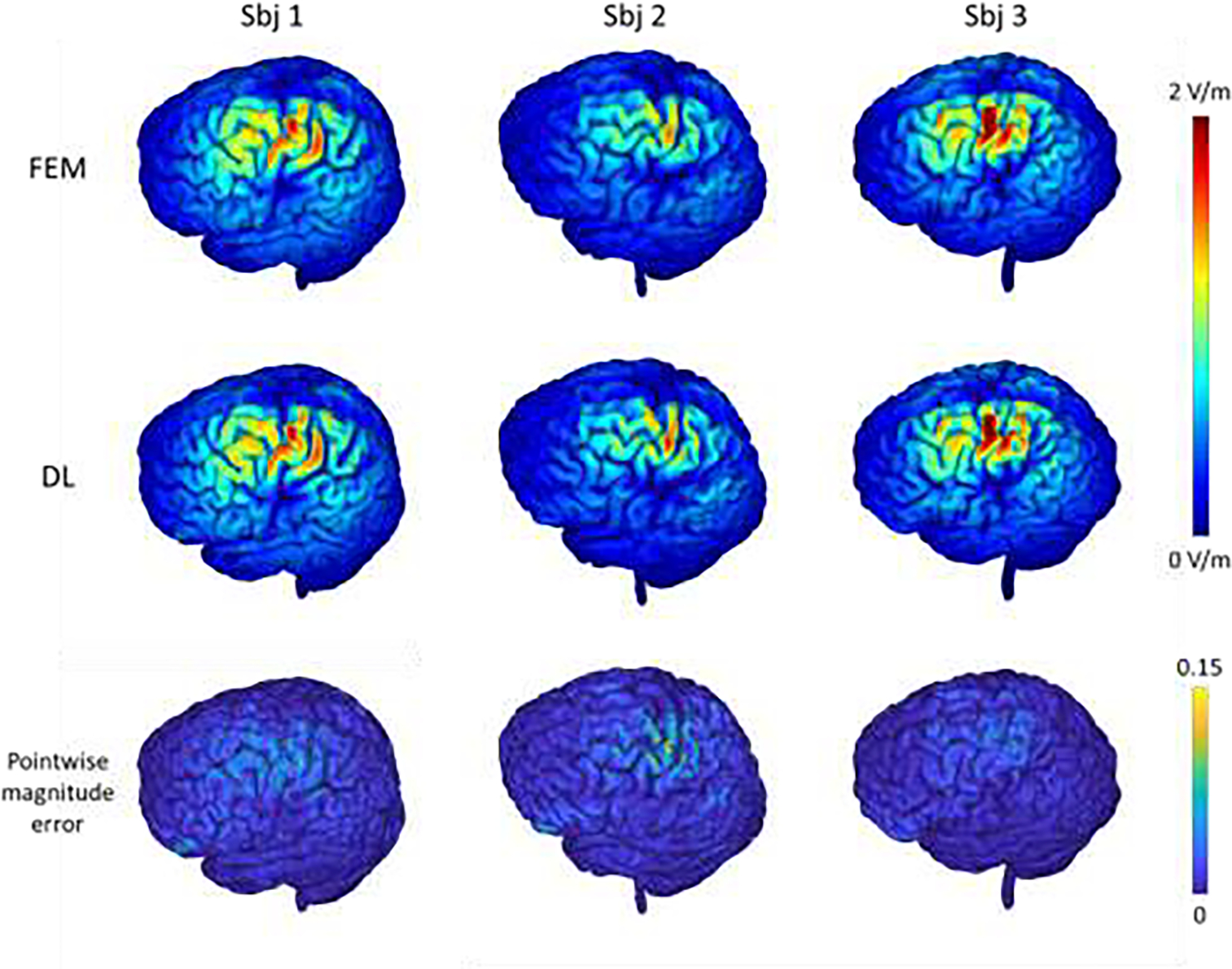
Fig.5.

E-fields of three randomly selected testing subjects computed by the FEM and the proposed DL model with the motor cortex as a stimulation target (1st and 2nd rows), and the corresponding pointwise magnitude error map with the FEM solution as reference (3rd row). The top colorbar shows the magnitude values (in V/m) of the E-fields and the bottom colorbar shows the normalized pointwise magnitude error.
