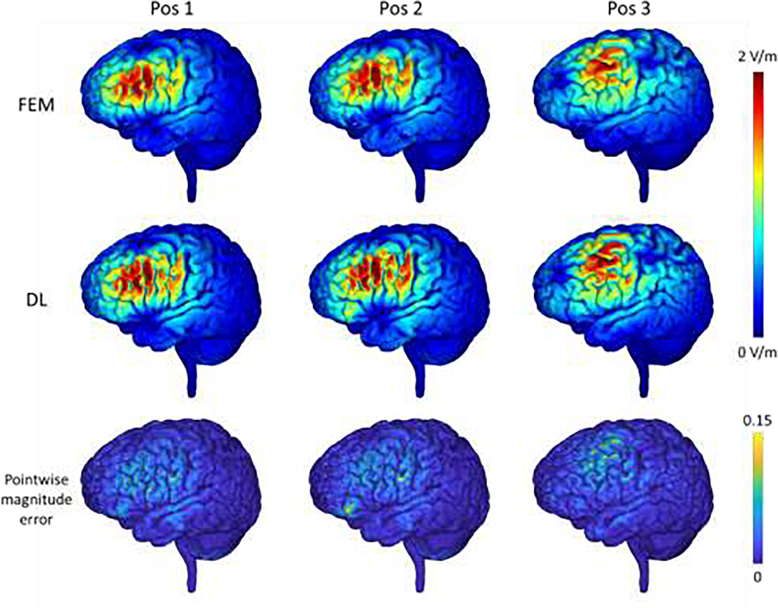
Fig.7.

E-fields of one randomly selected testing subjects computed by the FEM and the proposed DL model with the dorsolateral prefrontal cortex (DLPFC) as a stimulation target and the coil set at varying positions and in different directions (1st and 2nd rows), and the corresponding pointwise magnitude error map with the FEM solution as reference (3rd row). The top colorbar shows the magnitude values of the E-fields and the bottom colorbar shows the normalized pointwise magnitude error.
