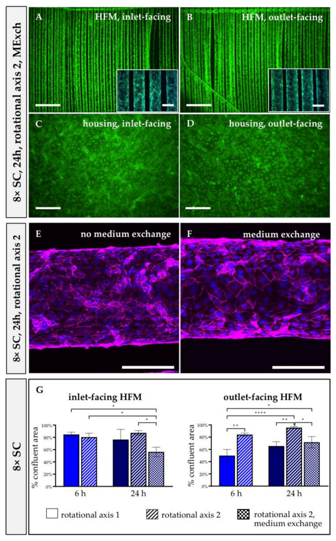
Figure 3.
Confluent EML on HFMs remained unaffected after medium exchange during 24 h rotational seeding. Following seeding procedure using 8 × SC for 24 h and RA2, calcein (green) and Hoechst 33342 (blue) staining was performed, and fluorescence microscopy images were taken from HFMs (A,B) and housing (C,D) either facing the inlet (A,C) or outlet (B,D). Scale: 2 mm, insert boxes in A, B, scale: 400 µm. (E,F) CLSM images for the detection of VE-cadherin (magenta) on HFM samples cultivated without (E) or with MExch (F). Nuclei were counterstained with Hoechst 33342 (blue). Scale: 200 µm. (G) Quantification of the successfully endothelialized HFM area per total available HFM surface. SC: seeding concentration. * p < 0.05, ** p < 0.01, **** p < 0.0001.

