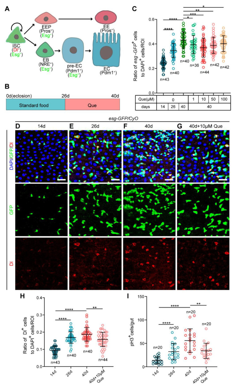
Figure 1.
Que prevents gut hyperplasia in aged Drosophila. (A) The Drosophila intestinal stem cell (ISC) lineages. ISCs (marked by Dl and Esg) divide asymmetrically to renew themselves and give rise to enteroendocrine progenitor cells (EEPs; marked by Esg and Pros) or enteroblasts (EBs; marked by Esg and NRE). EEPs further differentiate into enteroendocrine cells (EEs; marked by Pros), whereas EBs with high Notch signaling (pre-ECs; marked by Esg and Pdm1) further differentiate into enterocytes (ECs; marked by Pdm1). (B) Schematic diagram showing the process of feeding quercetin (Que) to Drosophila. (C) The ratio of esg-GFP+ cells to DAPI+ cells per region of interest (ROI) in the posterior midguts of 14-, 26-, and 40-day-old flies (esg-GFP/CyO) without Que supplementation and 40-day-old flies fed with four concentrations (1, 10, 50, and 100 µM) of Que. n: number of ROI counted. (D–G) Representative immunofluorescence images of 14- (D), 26- (E), and 40-day-old (F) posterior midguts without Que supplementation and 40-day-old posterior midguts with 10 µM Que supplementation (G) stained with DAPI (blue; nuclei), GFP (green; ISCs and progenitor cells marker), and Dl (red; ISCs marker). The top panels represent the merged images, the middle panels represent esg-GFP, and the bottom panels represent Dl. Scale bars represent 25 µm. (H) The ratio of Dl+ cells to DAPI+ cells per ROI in the posterior midguts of flies in experiments D-G. n: number of ROI counted. (I) The number of pH3+ cells in the whole guts of flies in experiments D-G. n: number of guts counted. Error bars represent standard deviation (SDs). Student’s t-tests, * p < 0.05, ** p < 0.01, *** p < 0.001, and **** p < 0.0001.

